


Classification and Application of Ultrasound Probes
During ultrasound examinations, the transmission and reception of ultrasound waves are both achieved through the transducer. The performance of the transducer directly affects the characteristics of the ultrasound waves and the quality of ultrasound imaging. Different transducers are suitable for different scanning areas; let’s learn more about them.
Classification and Application of Ultrasound Probes
Ultrasound probes come in various models. By diagnostic area, they can be classified into cardiac, abdominal, superficial organs, and endocavitary probes; by application method, into intracorporeal, extracorporeal, and biopsy-guidance probes; by the number of transducer elements used in the probe, into single-element and multi-element probes; and by beam steering method, into linear array, phased array, mechanical sector, and volumetric (matrix array) probes.
There are three most commonly used ultrasound probes for bedside point-of-care ultrasound examinations: linear array probe, phased array probe, and convex array probe.
01
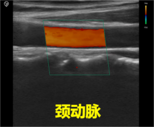
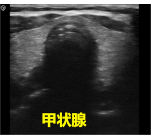
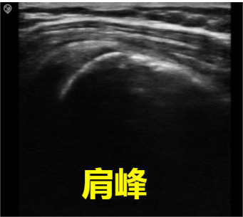
Linear array
Common frequency range: 5.0MHz-16.0MHz,
Common scan width: 30-40mm,
Imaging depth: within 6cm from the skin.
Features: The probe surface is flat, with a large contact area, rectangular imaging field of view, high imaging resolution, and relatively low penetration depth; suitable for examination of superficial vessels, small organs, musculoskeletal structures, etc.
02
phased array
Common frequency range: 1.5MHz-4.5MHz
Imaging depth: up to 35cm from the skin.
Features: The probe surface is flat with a small contact area. It can be scanned between ribs, with the smallest near-field field of view, a large far-field field of view, and a fan-shaped imaging field of view, suitable for the heart.
03
convex array
Common frequency range: 2.0MHz-5.0MHz
Common radius of curvature: 50-60mm
Imaging depth: within 30cm from the skin.
Features: The probe surface is convex, the imaging field of view is fan-shaped, with a large far-field field of view, and due to the relatively low frequency, it has higher penetration. It is widely used in abdominal, obstetric, and gynecological examinations, as well as other deep-seated organs.
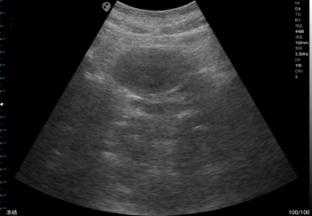
bladder
Probe maintenance and care
The effective accuracy of the ultrasound probe directly affects the efficiency of ultrasound examinations, and daily use and maintenance are crucial.
Ultrasound probes are expensive devices and should be handled gently to avoid impacts that may damage the internal crystals. Using non-corrosive coupling gel can extend their service life. Additionally, since ultrasound probes frequently come into direct contact with patients’ skin or mucous membranes, proper cleaning and disinfection are crucial. Disinfection should be performed according to the manufacturer’s instructions based on the examination site and intended use to prevent cross-infection.