


Fundamentals of Ultrasound: Acoustic Properties of Human Tissues
Most human organs can be examined using medical ultrasound, including common examples such as superficial organs (thyroid, breast, salivary glands, superficial lymph nodes, etc.), the heart and blood vessels, abdominal organs (liver, spleen, gallbladder, pancreas, adrenal glands, etc.), the genitourinary system (kidneys, ureters, bladder, uterus, ovaries, scrotum, testes, etc.), skin and muscle tissues, brain tissue, and more. To understand how different tissues and organs appear on ultrasound imaging, let’s explore the details.
01
Internationally, the intensity of human tissue reflection echoes is classified into four grades: high echo, isoecho, hypoecho, and anechoic. For more detailed description, it can also be further divided into high echo, relatively high echo, isoecho, hypoecho, relatively hypoecho, and anechoic. High echo accompanied by a distinct acoustic shadow posteriorly may also be referred to as strong echo.
1. Hyperechoic: High echogenicity is produced by bones, calculi, gas, fibrous connective tissue, etc. A acoustic shadow is often present behind bones and calculi.
2. Isoechoic: The echogenicity intensity of normal liver parenchyma is defined as isoechoic.
3. Hypoechoic: The echogenicity intensity is lower than that of normal liver parenchyma.
4. Anechoic: Normal human blood, urine, etc., appear as anechoic, and pathological changes such as effusions, cysts, abscesses, etc., also appear as anechoic.
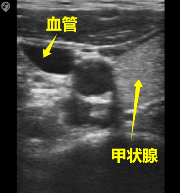
SonoMaxx Handheld Ultrasound MX6 for Thyroid and Thyroid Cyst Scanning
Overall, liquid structures appear as anechoic dark areas, while solid structures exhibit varying degrees of echogenicity. Homogeneous solid structures present as uniformly hypoechoic or isoechoic. Heterogeneous structures demonstrate mixed echogenicity. Calcified or gas-containing structures exhibit extremely strong echogenicity with posterior acoustic shadows.
It should be noted that the terms “hyperechoic,” “isoechoic,” and “hypoechoic” in sonographic imaging are relative. In most cases, these classifications are determined by comparison with the echogenicity of the observed tissue or organ. Additionally, they are influenced by the performance and settings of the imaging equipment, such as probe frequency, dynamic range, gain, and the use of tissue harmonics.
02
The sound attenuation of different tissues in human body is as follows: bone, calcification, stone> scar, cartilage, tendon> liver, kidney, muscle, brain> fat, blood> urine, bile, cyst fluid, pleural effusion, ascites.
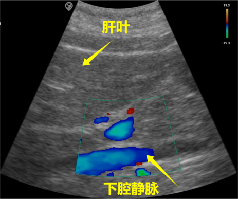
SonoMaxx Handheld Ultrasound MX6 for Inferior Vena Cava Scanning
Bones, calcifications, calculi, and gas-filled lungs exhibit strong echogenicity with posterior acoustic shadows. However, certain calculi and calcified structures, such as small nodules and minor calcifications, may lack acoustic shadows due to factors including ultrasound frequency, focal point, and incident angle.
Solid organs such as the liver, kidney, spleen, and pancreas exhibit isoechoic properties, with variations in echo intensity among different solid organs.
Fat is hypoechoic, but when the fat tissue forms an interface with other tissues, it appears hyperechoic. Urine, bile, cyst fluid, pleural effusion, and ascites (transudate) are anechoic.
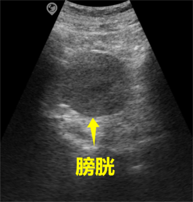
SonoMaxx Handheld Ultrasound MX6 for Bladder Scanning
The acoustic properties of pathological tissues can be classified into liquid, solid, calcified, and gas. The acoustic characteristics of the same disease may vary at different stages of its course, with corresponding differences in echogenicity.