Fundamentals of Ultrasound: Parameter Adjustment of Ultrasound Images
Common parameter adjustment for B mode
Gain
The degree of increase in the intensity of ultrasonic echo signal is visually represented as the brightness of the image on the ultrasound image.
Increasing the gain makes the image brighter, presenting more information, but also increasing the noise;
decreasing the gain makes the image darker, presenting insufficient information, but suppressing the noise.
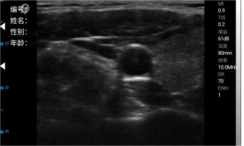
Too low
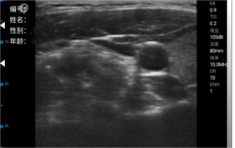
too high
The focus aims to converge and narrow the beam emitted by the probe, thereby enhancing the lateral resolution of the acoustic beam. The focal point is typically placed in the region of interest. An increased number of focal points can improve the overall lateral resolution of the acoustic field, but it significantly reduces the frame rate, making it unsuitable for observing rapidly moving organs.
Deep depth adjustment: The image will be reduced in size, affecting observation and diagnosis;
shallow depth adjustment: The image will be enlarged, with a corresponding decrease in resolution.
The appropriate depth should be selected based on the proximity of the observed tissue, organ, or lesion to the mid-field of the image area.
Dynamic range
Reducing the dynamic range enhances image contrast and sharpness, but it diminishes the display of useful information, impairing observation and judgment.
Increasing dynamic range: Reducing image contrast enhances image detail, though it provides more information. However, decreased contrast may impair the observation of certain lesions and result in indistinct boundaries between tissues and lesions.
Common parameter adjustment for C mode
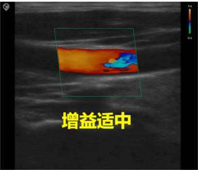
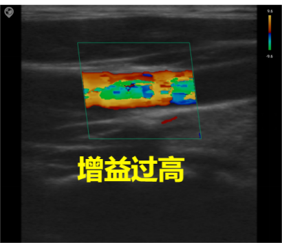
Gain
Total gain: Adjusts the sensitivity of blood flow signals, with the gain value displayed in real-time on the image parameter area of the screen.
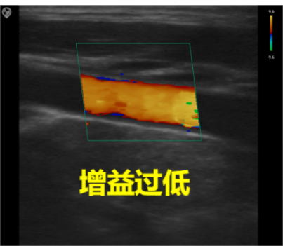
Image quality: Excessive gain obscures the image with chaotic color spots, while insufficient gain may lead to loss of blood flow signals. During actual adjustment, the optimal setting is to clearly distinguish red and blue blood flow without color aliasing.
PRF – Pulse Repetition Frequency
Sampling Frame
Sampling frame size: A smaller sampling frame results in a higher frame rate, while a larger sampling frame can display more blood flow information. The sampling frame size is clinically adjusted according to actual needs.
Sampling frame deflection angle: Generally, clinical adjustments are made by slightly deflecting the sampling frame to obtain better blood flow visualization.
Common parameter adjustment in PW mode
Angle of deflection
Sampling volume
The excessive sampling volume results in the acquisition of a large amount of intravascular blood flow signals.The non-blood flow motion interference signal increased, and the spectrum was wide.
The volume of blood sample is too small, the velocity distribution range of red blood cells contained in the sample is reduced, and the spectrum is too narrow.
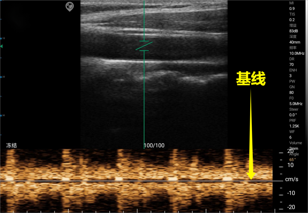
Baseline