


Fundamentals of Ultrasound Principles of Ultrasound Imaging
Ultrasound examination is non-invasive, painless, and radiation-free, with low cost, and has gradually developed into one of the essential diagnostic methods in clinical practice. To help physicians better understand and master ultrasound technology, the “Palm Vision” Eye Academy’s ultrasound fundamentals column is now live!
More learning columns are coming soon. Stay tuned!
Principles of Ultrasonic Imaging
Ultrasound imaging utilizes the probe to emit ultrasonic waves, which scan the human body. Due to the varying acoustic impedance of tissues, reflected ultrasonic waves are generated. The probe receives the reflected ultrasonic waves and converts them into electrical signals, which are then processed by the system to display as different images.
What is ultrasound
The audible frequency range of human ears is 20-20000Hz, and sound waves beyond this range are generally referred to as ultrasonic waves.
Basic Concepts of Ultrasonic Waves
An object capable of producing sound is termed a sound source, with its fundamental origin being vibration. In clinical diagnostics, the probe’s crystal chip serves as the sound source.
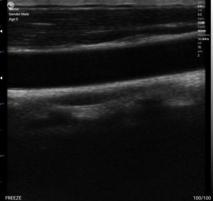
Propagation characteristics of ultrasonic waves
The speed of ultrasound varies with the medium. The speed of ultrasound in human soft tissue is about 1540 m/s.
Meanwhile, ultrasonic propagation is also influenced by the density and hardness of the medium, which is referred to as acoustic characteristic impedance in physics. The acoustic impedance value of different media is the product of their density and sound velocity. Simply put, it represents the tissue’s resistance to ultrasonic waves, with stronger resistance leading to more pronounced reflection.

– Primary Care Clinics / Community Hospitals: SonoMaxx MX9 Ultra (Host + 2 core probes, total budget within 30,000 CNY, covering all needs)
– High-End Private Clinics: Butterfly iQ+ + Clarius PA HD3 (Focus on technological advantages and specialist diagnosis capabilities)
– Mobile Medical Services / Veterinary Institutions: Youkey D8 (Long battery life) + Dr. Sono Convex Pro (High cost-effectiveness)
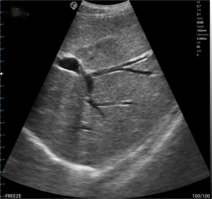
In sonograms, various echo imaging patterns are primarily caused by differences in acoustic impedance. As shown in the figure above, due to the high acoustic impedance of bones, they appear as high echoes on the image. The general order of acoustic impedance values is: Air <Fat <Water <Blood <Kidney <Liver <Muscle <Skin <Bone