Fundamentals of Ultrasound: Ultrasonic Artifacts

Ultrasound artifacts are particularly significant in ultrasound diagnosis. Identifying these artifacts serves dual purposes: it prevents misdiagnosis or missed diagnosis caused by artifacts, while leveraging certain characteristics of the artifacts can enhance the detection of specific pathological conditions or components. Today, let us explore ultrasound artifacts in detail.

01

What is an ultrasound artifact?

Ultrasound artifacts refer to the discrepancy between the ultrasonographic cross-sectional images and the actual anatomical structures, also known as ultrasound artifacts, artifacts, or false images. These manifest as additions, reductions, or distortions of echo information in the sonographic images.
Ultrasound examination is characterized by a significant number of artifacts. These artifacts exhibit universality, polymorphism, developmental nature, and multiplicity. The process of ultrasound diagnosis is markedly dependent on manual techniques, making it nearly impossible to completely eliminate artifacts. Simultaneously, for physicians, artifacts serve a dual role as both interference and guidance.

02

Common ultrasound artifacts

The main sources of artifacts are improper scanning techniques and inherent physical limitations in ultrasound propagation. 

Common types of ultrasound artifacts include the following:

Multiple reflection artifact

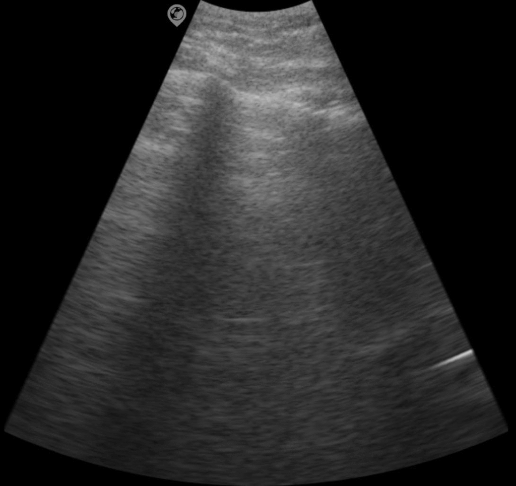
During sound beam propagation, an interface parallel to the transducer is encountered, where significant differences in acoustic impedance on both sides of the interface generate strong reflected waves. These reflected waves undergo multiple reflections between the transducer and the interface, forming a series of repeatedly distorted and sequentially distorted reflection images. This phenomenon is commonly observed in structures such as the lungs and trachea.

The image shows the multiple reflection artifact of the lung

Enhanced posterior echo

When the ultrasonic beam passes through fluid-filled organs or lesions, the resulting attenuation is compensated by excess, leading to enhanced posterior echoes. This phenomenon is commonly observed during the scanning of cysts, bladder, and similar structures.

Acoustic shadow

During ultrasonic propagation, strong reflections occur when encountering interfaces with significantly different acoustic impedances, such as stone-water or gas-soft tissue interfaces, resulting in pronounced acoustic attenuation behind these interfaces. This phenomenon is commonly observed behind bones, calculi, and calcified foci.
The image shows the sonogram of the flexor pollicis longus tendon

Side lobe artifact

The acoustic beam emitted by a sound source has a primary lobe, typically centered at the source’s axis perpendicular to its surface, known as the main lobe. Around the main lobe are symmetrically distributed pairs of smaller lobes called side lobes. During scanning imaging, the side lobes are simultaneously captured. Side lobe artifacts occur when the far-field side lobe echoes of the probe are excessively strong and overlap with the main lobe echoes, commonly observed in areas such as the uterus, gallbladder, and diaphragm.

Mirror artifact

When ultrasound is projected onto a large, smooth, and hyperechoic interface, it produces a symmetrical reflection phenomenon similar to that of a plane mirror, which is caused by signal delay. This is commonly observed near the diaphragm. A solid tumor or fluid-filled space-occupying lesion may be displayed on both sides of the diaphragm. The area closer to the diaphragm appears as a solid shadow, while the deeper area appears as a shadow or mirror image.

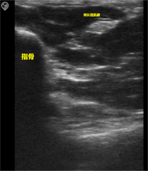
Aartial volume effect artifact
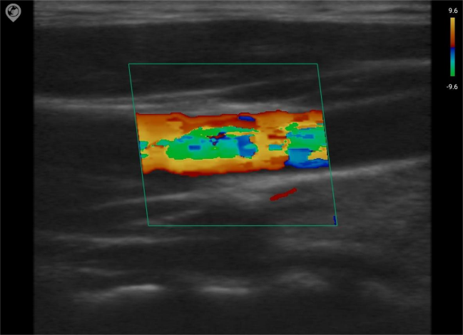
Due to resolution limitations, the echo signal actually covers information of a certain volume of space, and the echoes of the lesion and the surrounding tissues may overlap and be displayed in the same image, resulting in interference with the imaging of the lesion.

Aliasing

In Doppler ultrasound, when the blood flow velocity exceeds the upper limit determined by the Nyquist sampling theorem, the measured velocity reverses in sign, causing the Doppler flow color to change from red-yellow to blue.
Color Blood Flow Mode of Carotid Artery Measurement with SonoMaxx Handheld Ultrasound MX6

In summary, the identification of ultrasound artifacts requires extensive knowledge and proficient operational skills. It is essential to promptly eliminate interference artifacts, scientifically utilize indicator artifacts, and make appropriate diagnoses to better apply ultrasound in clinical practice.

Leave a Reply

Your email address will not be published. Required fields are marked *