Fundamentals of Ultrasound: Ultrasound Imaging Modes
In the 1950s, Type A and Type M ultrasound were introduced; in the 1970s, Type B ultrasound (commonly known as “B-mode ultrasound”) gradually emerged; in the 1980s, color Doppler ultrasound (colloquially referred to as “color ultrasound”) was developed. Over the past three decades, medical ultrasound diagnostic technology has undergone successive revolutionary leaps and has now become the preferred method for diagnosing various clinical conditions. So, what are the modes of ultrasound?
Amplitude
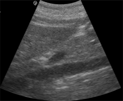
Brightness
Motion
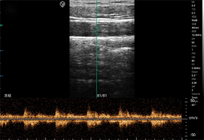
The M-mode displays the intensity of tissue echo signals based on brightness levels, while also showing the movement trajectories of these light spots on the time axis, reflecting one-dimensional tissue structure and motion information.
M-mode ultrasound is primarily employed for the examination of the cardiovascular system, analyzing the motion amplitude of the heart and major blood vessels, dynamically assessing the morphological structure and functional status of the cardiovascular system, and obtaining corresponding physiological or pathological technical indicators of the vessels.
color
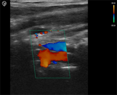
Doppler