Description
SonoMaxx MX7 Wireless Handheld Color Doppler Ultrasound Scanner is a groundbreaking point-of-care (POC) diagnostic tool engineered by SonoMaxx, designed to redefine portability, performance, and versatility in whole-body imaging. As part of BMV’s MX Scanners family (alongside the MX9), the MX7 combines ultra-lightweight design (220g), powerful imaging technology, and seamless connectivity to empower clinicians to perform high-quality scans anytime, anywhere—from hospital wards and emergency rooms to remote clinics and veterinary practices. With 6 specialized transducers, advanced imaging features, and intuitive operation, it delivers crystal-clear images and comprehensive clinical insights, making it an indispensable tool for primary care, critical care, specialty medicine, and veterinary care.
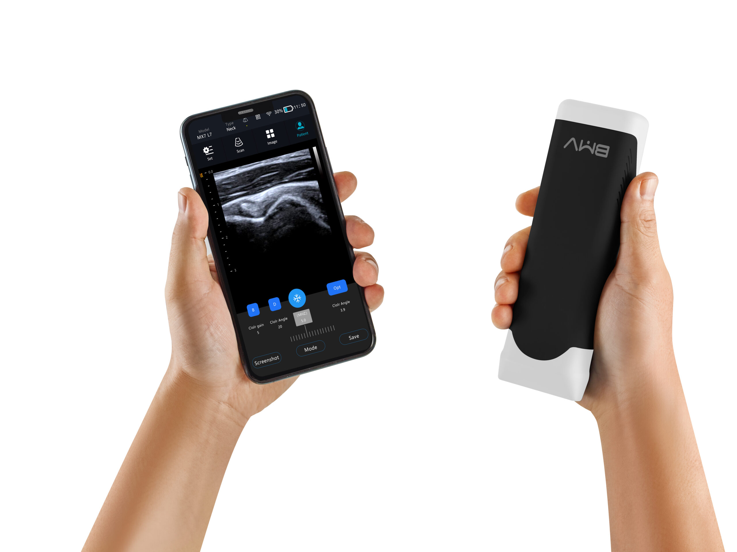
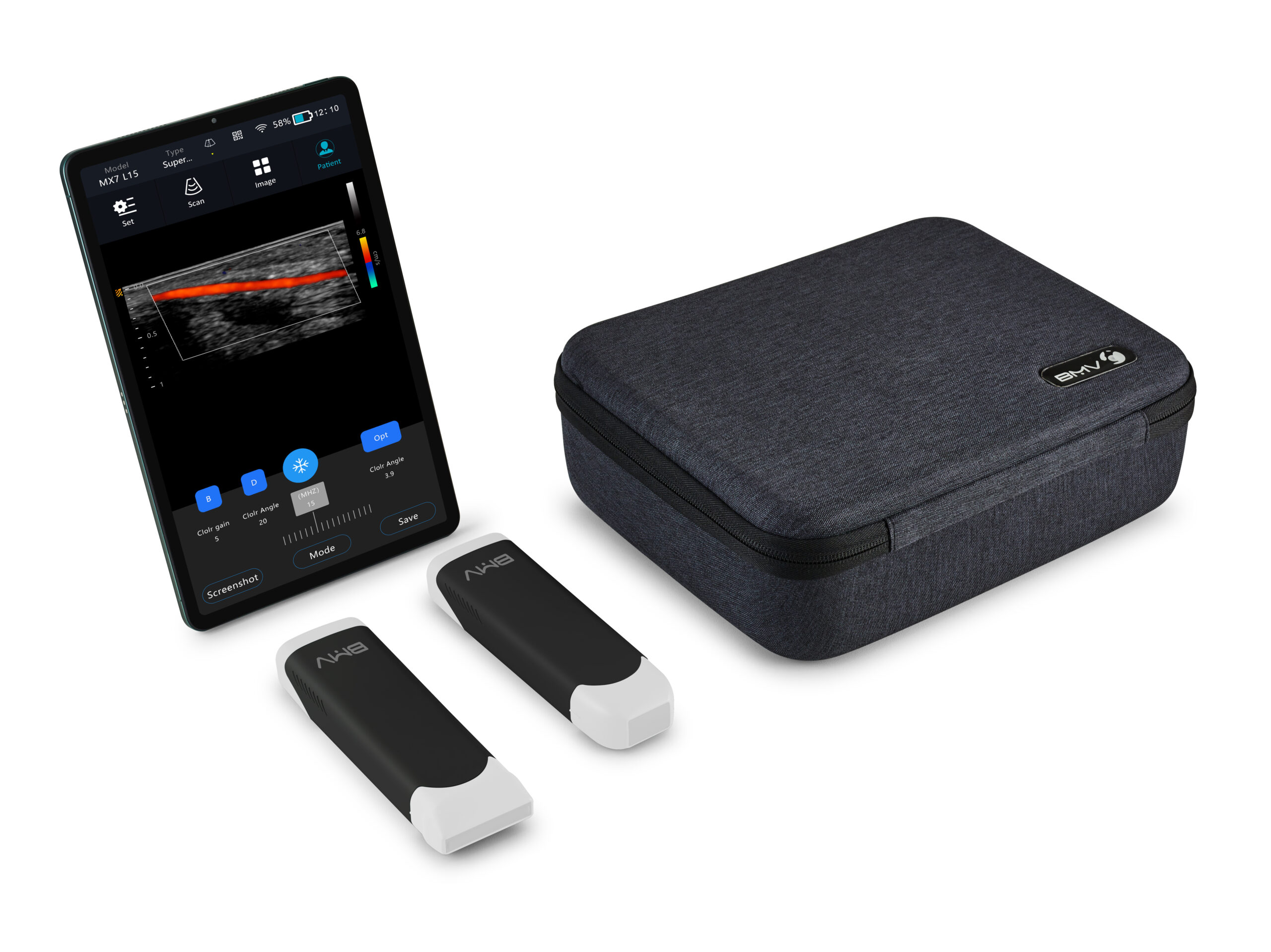
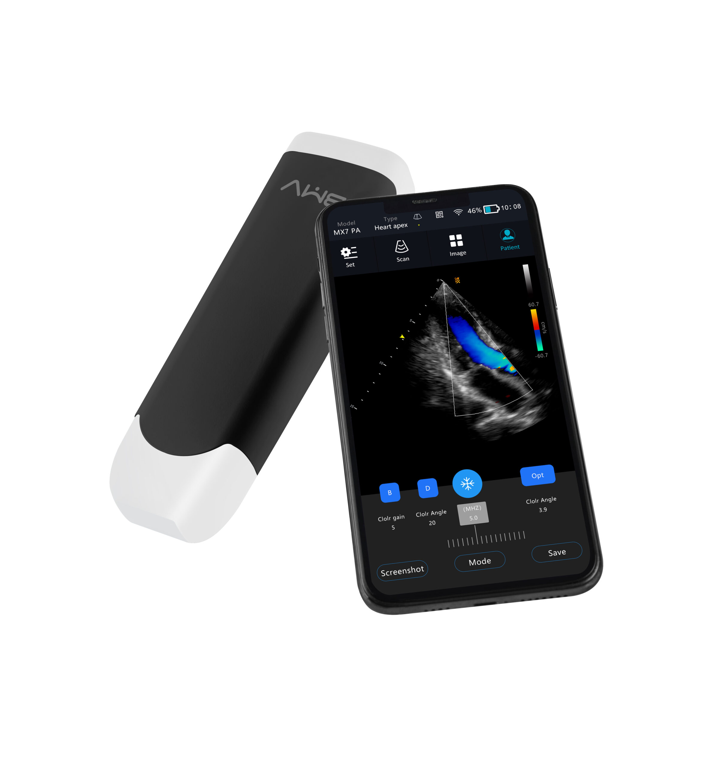
At the core of the MX7’s excellence is its robust hardware and advanced imaging capabilities. Equipped with 64 physical channels and 192 piezoelectric elements, it produces high-resolution, smooth images with 256 scan lines and supports a full suite of imaging modes: B, C (Color Doppler), M, PW (Pulsed Wave), PD (Power Doppler), and DPD (Dual Power Doppler). Key enhancements include Harmonic Imaging (for higher image quality and reduced artifacts) and Enhanced Needle Visualization (ENV)—a critical feature for ultrasound-guided procedures that improves needle visibility even at steep insertion angles. The system also offers PW Automatic Measurement, generating 13 parameters and 34 indicators for carotid artery assessments—essential for screening atherosclerosis in elderly patients.
Portability is a hallmark of the MX7: weighing only 220g (≤250g) with compact dimensions (188×75×34mm), it fits easily in a coat pocket or medical bag, enabling fatigue-free use across multiple patients. Its one-hand user interface and instant startup (just flip the top) streamline workflow, while preconfigured presets for common applications integrate seamlessly into clinical routines. A built-in rechargeable battery supports out-of-hospital use, ensuring reliability in mobile or power-limited settings.
The MX7 offers 6 specialized transducers to cover whole-body imaging needs, with smart identification for carotid artery (CCA) and Intima-Media Thickness (IMT) measurement:
- PA HD (Phased Array): 1–5 MHz, 18.5cm max depth—ideal for bedside cardiac, FAST, abdominal, bladder, lung, OB/GYN, and superficial vascular access.
- C2 HD (Convex): 2–5 MHz, 38cm max depth—specialized for abdominal, lung, and OB/GYN imaging.
- C5 HD (Microconvex): 2–5 MHz, 31cm max depth—suited for small parts, MSK, pediatrics, veterinary care, and abdominal/cardiac/lung imaging.
- C7 HD (Microconvex): 5–8 MHz, 12cm max depth—optimized for small parts, MSK, pediatrics, speech therapy, and veterinary applications.
- L7 HD (Linear): 6–11 MHz, 12cm max depth—designed for vascular, lung, breast, MSK, nerve, ocular, small parts, animal, and anesthesia imaging.
- L15 HD (High-Frequency Linear): 6–15 MHz, 10cm max depth—perfect for injections, nerve blocks, breast, MSK, thyroid, small parts, lung, and vascular imaging.
Connectivity and collaboration are seamless with 5G WiFi support, enabling zero-latency data transfer, remote diagnosis, and education via SonoMaxx’s optional Remote Diagnosis and Training Platform. The scanner stores images in JPG/PNG/BMP/DCM (DICOM) formats, with cineloop functionality (100 frames) for retrospective review. Comprehensive measurement tools include area, bore narrow, ellipse, distance, angle, and hip joint calculations, while needle guidance adapts to 14-22G needles with preset puncture angles (18°/25°/35° for convex probes; 42°/52°/62° for linear probes) for safe, precise interventional procedures.
Built for durability and clinical practicality, the MX7 features GUP Enhanced Processing for improved image quality and zero latency, ensuring real-time visualization of anatomical structures and blood flow. Its user-friendly design, combined with powerful clinical capabilities, makes it suitable for clinicians of all experience levels—from seasoned specialists to primary care providers. Whether used for cardiac assessments, vascular screening, MSK imaging, or veterinary care, the SonoMaxx MX7 delivers hospital-grade performance in a handheld form, proving that advanced diagnostics can be both portable and accessible.
Core Product Advantages
1. Ultra-Portable & User-Friendly Design
- 220g Ultra-Lightweight: Compact dimensions (188×75×34mm) for fatigue-free extended use and easy transport.
- Instant Startup: Flip-top design for quick activation, fitting seamlessly into busy clinical workflows.
- One-Hand Operation: Intuitive interface and presets for common applications reduce learning curves.
- Built-In Battery: Supports out-of-hospital use, ideal for mobile and remote care.
2. Powerful Imaging & Advanced Clinical Features
- 64 Channels + 192Elements: Delivers high-resolution, smooth images with 256 scan lines.
- Comprehensive Modes: B/C/M/PW/PD/DPD + Harmonic Imaging for reduced artifacts and enhanced contrast.
- Enhanced Needle Visualization (ENV): Improves needle visibility at steep angles, supporting safe guided procedures.
- PW Automatic Measurement: Generates 13 parameters + 34 indicators for carotid artery screening (atherosclerosis detection).
3. 6 Transducers for Whole-Body Coverage
- Diverse Probe Lineup: Phased array, convex, microconvex, linear, and high-frequency linear transducers cover cardiac, abdominal, vascular, MSK, OB/GYN, pediatric, and veterinary needs.
- Smart Identification: CCA and IMT measurement for vascular health assessments.
- Wide Depth Range: 10cm–38cm max depth, adapting to superficial and deep tissue imaging.
4. Seamless Connectivity & Collaboration
- 5G WiFi Support: Zero-latency data transfer, remote diagnosis, and education.
- Remote Platform Compatible: Optional Remote Diagnosis and Training Platform for global collaboration.
- DICOM Compatibility: Stores images in DCM format for integration with hospital PACS systems.
5. Precise Needle Guidance & Measurement Tools
- Needle Adaptability: Supports 14-22G needles with preset puncture angles for convex/linear probes.
- Comprehensive Measurements: Area, distance, angle, hip joint, and vascular parameters for accurate diagnostics.
- Cineloop Function: 100-frame review for detailed analysis of dynamic structures.
| Specification Category | Parameter Details |
| Product Model | SonoMaxx MX7 Wireless Handheld Color Doppler Ultrasound Scanner |
| Core Configuration | 64 physical channels; 192 piezoelectric elements; 256 scan lines |
| Imaging Modes | B, Color Doppler (C), M, Pulsed Wave (PW), Power Doppler (PD), Dual Power Doppler (DPD); Harmonic Imaging |
| Transducer Specifications | 1. PA HD (Phased Array): 1–5 MHz, Max Depth 18.5cm (cardiac, FAST, abdominal, OB/GYN)2. C2 HD (Convex): 2–5 MHz, Max Depth 38cm (abdominal, lung, OB/GYN)3. C5 HD (Microconvex): 2–5 MHz, Max Depth 31cm (pediatrics, MSK, veterinary)4. C7 HD (Microconvex): 5–8 MHz, Max Depth 12cm (small parts, speech therapy, veterinary)5. L7 HD (Linear): 6–11 MHz, Max Depth 12cm (vascular, MSK, anesthesia)6. L15 HD (High-Frequency Linear): 6–15 MHz, Max Depth 10cm (nerve, thyroid, small parts) |
| Key Features | Enhanced Needle Visualization (ENV); PW Automatic Measurement (13 parameters/34 indicators); IMT Measurement; Smart CCA Identification; Zero Latency; GUP Enhanced Processing |
| Needle Guidance | Convex Probe: 18°/25°/35° puncture angles; Linear Probe: 42°/52°/62° puncture angles; Adaptive Needle Type: 14-22G |
| Display & Operation | One-hand user interface; Instant startup (flip-top design); Presets for common applications |
| Connectivity | 5G WiFi; Optional Remote Diagnosis and Training Platform |
| Data Management | Image Formats: JPG/PNG/BMP/DCM (DICOM); Cineloop: 100 frames |
| Measurement Tools | Area, bore narrow, ellipse, distance, angle, hip joint calculations |
| Physical Specifications | Dimensions: 188mm×75mm×34mm; Net Weight: 220g (≤250g) |
| Power & Usage | Built-in rechargeable battery; Suitable for in-hospital and out-of-hospital use |
| Supported Specialties | Cardiology, Vascular, MSK, OB/GYN, Pediatrics, Emergency Medicine, Anesthesia, Veterinary, Primary Care, Critical Care |